Identifying Unknown Strains Using 16S rRNA Gene Sequencing
This blog provides a detailed, step-by-step guide to identifying and characterizing unknown bacterial strains using 16S rRNA gene sequencing. It begins with the collection of samples from various environments such as soil, air, and water, followed by bacterial strain isolation and growth characterization. The blog then explains key techniques like Gram staining, DNA extraction, and PCR amplification of the 16S rRNA gene. The focus shifts to the quality check of DNA using gel electrophoresis and fluorometric methods, before moving on to Sanger sequencing. Finally, it discusses the analysis of sequencing data to accurately identify and classify the bacterial strain, offering a comprehensive overview of molecular microbial identification.
![]() Ankita Shastri
Feb 28, 2025 05:14
2019
1
Ankita Shastri
Feb 28, 2025 05:14
2019
1

Decoding Bacterial Mysteries: A Comprehensive Guide to Identifying Unknown Strains Using 16S rRNA Gene Sequencing
In molecular biology, the identification and characterization of unidentified bacterial strains are essential procedures. One of the most powerful and widely used techniques for bacterial identification is 16S rRNA gene sequencing. This technique makes use of the 16S ribosomal RNA gene's high degree of conservation among bacterial species, which makes it a perfect target for bacterial organism identification and classification. The procedures for identifying and characterizing an unidentified bacterial strain using 16S rRNA gene sequencing will be covered in this blog.
1. Sample Selection from Various Sources like Soil, Air, Water, etc.
The first step in identifying an unknown bacterial strain is to carefully select a sample from an environment where bacteria are likely to be abundant. There is a lot of microbial diversity in nature, and isolating the target microorganisms requires careful selection of the sample source. The type of bacteria being studied determines the source to use, as different habitats offer distinct microbial communities. For example:
- Air samples often contain bacteria from plant matter, human activity, and environmental exposure. As members of the bioaerosol community, airborne bacteria can be investigated to learn more about air quality, microbial dispersal, and their impact on human health. Filtration or impaction techniques are frequently used in air sampling procedures.
- Soil samples are rich in microorganisms due to the high availability of nutrients and organic matter. Numerous types of bacteria are frequently found in soil samples, particularly those that are involved in the cycling of nutrients, including decomposers or nitrogen-fixing bacteria. The potential uses of soil microorganisms in bioremediation make them interesting as well.
- Water samples may contain a variety of microorganisms, ranging from harmless bacteria to potentially pathogenic strains. Bacteria are abundant in aquatic bodies, including freshwater and marine settings. Identification of species involved in nutrient cycling, water purification, and even harmful bacteria is aided by sampling from various sources. Bacterial colonies are frequently isolated using methods like membrane filtration.
Proper sample collection is essential to avoid contamination, and the sample should ideally be processed as quickly as possible to prevent changes in the microbial composition.
2. Bacterial Strain Selection and Growth Characteristics
After collecting the sample, the next step is to isolate the bacterial strain. This can be done using standard microbiological techniques, such as-
- Streak Plate Method: A small volume of the sample is streaked on an agar plate, allowing individual bacterial cells to grow into isolated colonies.
- Serial Dilution: In cases of highly concentrated samples, serial dilution followed by plating allows for the growth of individual colonies from a diluted sample.
After separation, bacteria are grown under specific growth conditions, including temperature, pH and nutrient medium, helping to identify optimal bacterial growth parameters. Growth characteristics such as:
- Colony morphology: Colony appearance in terms of size, shape, color and texture.
- Growth rates: doubling time (generation time) and the time for achieving the exponential stage of growth.
- Oxygen requirements: bacteria reaction to aerobic, anaerobic or microaerophilic conditions. These characteristics give the first advice for the identity of the strain, but often not enough for precise identification.
These characteristics provide preliminary insights into the nature of the bacterium.
3. Growth Parameters, Gram Staining, Identification, and Characterization
To further understand the bacterial strain, it is important to assess its basic morphological and biochemical properties. This includes:
- Gram Staining: This staining technique differentiates bacteria into two groups—Gram-positive (purple) and Gram-negative (pink)—based on the structure of their cell walls. This provides crucial information about the bacterial cell wall composition and helps narrow down the species.
- Microscopic Examination: After Gram staining, the bacteria are observed under the microscope to determine their shape (e.g., cocci, bacilli, spirilla) and arrangement (e.g., chains, clusters, pairs).
- Biochemical Tests: Common tests include catalase activity, sugar fermentation, and motility assays. These tests help characterize the metabolic capabilities of the bacterial strain. A series of tests like this:
- Catalase Test: Distinguish preventive catalase tissue (e.g. Staphylococcus) from catalase-negative tissue (e.g. Streptococcus).
- Oxidase Test: Identifies bacteria that produce cytochrome using oxidases such as Pseudomonas species.
- Testing carbohydrates: Identify bacteria as a function of their ability to ferment sugar and produce acid or gas.
These parameters help reduce the types of bacteria, but molecular methods are necessary for the final identification.
4. Genomic DNA and Plasmid Isolation and Quantification
For molecular identification, the next step is to extract genomic DNA from the bacterial cells. This can be done using a variety of DNA extraction kits that break open the bacterial cells and separate the DNA from other cellular components. Additionally, plasmid DNA may also be isolated if the bacterium is known to contain plasmids.
The isolated DNA is then quantified, often using UV spectrophotometry at 260 nm or a fluorometric method like Qubit, which uses specific fluorophores to bind to DNA and provide more accurate quantification.
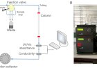
5. DNA Quality Check on Gel Electrophoresis
To ensure that the extracted genomic DNA is of high quality, a gel electrophoresis run is performed. Its integrity and quality must be verified. Electrophoresis of agarose gel gels is commonly used.
- DNA with high molecular weight: Good quality genomic DNA should look like separate, intact strips without degradation.
- Contaminants: RNA contamination can be assessed by the presence of RNA-compatible polymeric mass zones. DNA purity can be assessed by comparing absorption ratios at 260 nm and 280 nm (A260/A280).
Smearing or fragmented bands may indicate degraded DNA, which could affect downstream applications like PCR and sequencing.
6. DNA Quantification Using Fluorometric Method (Qubit)
After confirming the quality of the DNA, its concentration is measured using a fluorometric method like Qubit. The Qubit fluorometer uses a fluorescent dye that specifically binds to the DNA, allowing for precise quantification. This method provides more precise results than UV spectrophotometry by using fluorophores that bind to DNA, yielding a fluorescence signal proportional to DNA concentration. This accurate quantification is vital for ensuring optimal conditions for PCR amplification.
7. 16S rRNA Gene PCR Amplification Using Gene-Specific Primers
The next step in molecular identification is amplification of the 16S RNA gene using a chain reaction by polymerase (PCR). The 16S RNA gene represents approximately 1,500 basic and highly conservative pairs of bacteria, making it an ideal target for universal primers. PCR amplification involves:
- Primers: Universal primers targeting the conserved regions of the 16S rRNA gene (e.g., 27F and 1492R primers) are used to ensure broad amplification across bacterial species.
- Thermal Cycling: The reaction involves denaturation (to separate DNA strands), annealing (primers bind to the DNA), and extension (DNA polymerase synthesizes the complementary strand), typically performed in a thermocycler.
Successful amplification produces a PCR product that can be visualized on an agarose gel.
8. PCR Product Gel Extraction and Purification
After PCR amplification, the reaction products are analyzed by running them on an agarose gel. If the amplification is successful, a band corresponding to the 16S rRNA gene will appear. The next step is to extract and purify this PCR product using gel extraction kits, which remove any unwanted PCR components, such as primers or enzymes, to ensure high-quality DNA for sequencing.
9. Sanger Sequencing
The purified PCR product is then subjected to Sanger sequencing, a method based on chain termination. In this process:
- Fluorescently labelled dideoxynucleotides (ddNTPs) are incorporated into the growing DNA strand. These DDNTPs do not have 3\' hydroxyl groups that end in DNA synthesis for certain reasons.
- Capillary electrophoresis: Complete fragments are split by size and found using fluorescence to generate chromatograms corresponding to the nucleotide sequence.
Sanger sequencing is ideal for sequencing 16S RNA genes as it provides high accuracy and length length.
10. Sequencing Data-Based Taxonomy Analysis
The final step is to analyze the sequencing data to identify the bacterial strain. The obtained 16S rRNA gene sequence is compared against a reference database, such as NCBI GenBank or the Ribosomal Database Project (RDP). These databases contain a collection of 16S rRNA sequences from known bacterial species. By aligning the obtained sequence with those in the database, researchers can identify the closest matches and determine the taxonomic classification of the unknown strain.
The sequence identity percentage and phylogenetic analysis (using tools like BLAST or MEGA) will allow for precise classification to the genus or species level, providing a comprehensive identification of the bacterial strain.
11. Role of IBRI Noida in Molecular Biology Hands-on Training
This blog provides a detailed guide to identifying and characterizing unknown bacterial strains using 16S rRNA gene sequencing, covering processes from sample collection to sequencing data analysis. It highlights key steps like bacterial isolation, DNA extraction, PCR amplification, and Sanger sequencing, which are essential for accurate microbial identification. Institutes like Indian Biological Sciences and Research Institute (IBRI) Noida play a pivotal role in advancing molecular biology by offering specialized training in techniques such as PCR, sequencing, and bioinformatics. With state-of-the-art labs and expert guidance, IBRI Noida equips students and professionals with practical, hands-on skills, preparing them for real-world applications in microbial research and diagnostics.
Conclusion
16S rRNA gene sequencing is a powerful tool for the identification and characterization of unknown bacterial strains. The process involves multiple stages, each of which contributes to the accuracy and reliability of the results. From sample collection and strain isolation to PCR amplification, sequencing, and bioinformatics analysis, the 16S rRNA gene sequencing method provides a comprehensive framework for bacterial identification, enabling microbiologists to accurately identify and classify bacteria at the molecular level. This approach has applications in fields ranging from clinical diagnostics to environmental monitoring and biotechnological research.
Explore IBRI’s programs today to learn more about molecular biology trainings and unlock new career opportunities and stay ahead in the life sciences industry.
You can contact us at - Email: info@ibri.org.in or call at: (+91) 9999509892, 9971441910