MICROSCOPIC ANALYSIS OF BACTERIA IN HINDON RIVER WATER
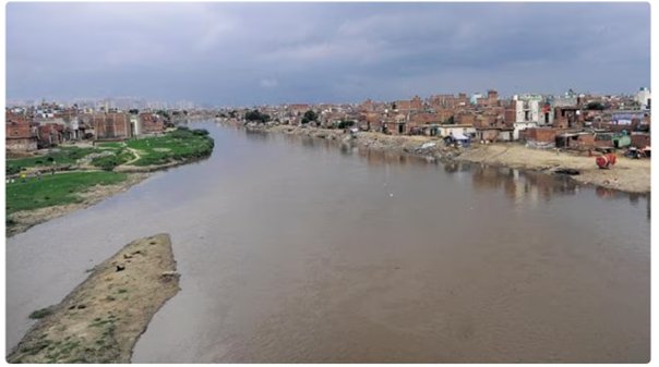
The Hindon River, a crucial water source, has been facing environmental challenges due to pollution and microbial contamination. Understanding the bacterial composition of river water is essential for assessing public health risks and ecological impact. This blog explores the microscopic identification of key bacterial species—Escherichia coli, Salmonella Typhi, Pseudomonas aeruginosa, and Bacillus subtilis—isolated from Hindon River water samples using Gram staining and morphological analysis. Through a detailed examination of cell structure, staining reactions, and environmental implications, this study highlights the presence of waterborne pathogens and their significance in ecosystem stability and disease transmission. The findings reinforce the importance of microbial surveillance for monitoring pollution levels, supporting sustainable water management, and ensuring the safety of natural water bodies.
![]() Ankita Shastri
May 16, 2025 12:02
2185
0
Ankita Shastri
May 16, 2025 12:02
2185
0
Introduction
Microbial analysis of water sources plays a crucial role in assessing water quality, pollution levels, and potential health hazards. Gram staining, a fundamental technique in microbiology, allows researchers to classify bacteria based on cell wall composition, aiding in the identification of waterborne pathogens.
This study focuses on Gram staining-based identification of bacteria isolated from Hindon River water samples, particularly Escherichia coli, Salmonella Typhi, Pseudomonas aeruginosa, and Bacillus subtilis—each linked to environmental contamination and public health concerns.
Principle of Gram Staining
Gram staining was developed by Hans Christian Gram (1884) and is one of the most widely used techniques in microbiology. It differentiates bacteria based on the ability of their cell wall to retain crystal violet dye after alcohol decolorization.
- Gram-positive bacteria have thick peptidoglycan layers, trapping crystal violet inside the cell. They appear purple after staining.
- Gram-negative bacteria possess thin peptidoglycan layers and an outer membrane, which loses crystal violet when exposed to alcohol. They take up safranin counterstain, appearing pink/red.
This difference has biological significance, as Gram-negative bacteria tend to be more resistant to antibiotics, given their protective outer membrane.
Materials and Methods
Sample Collection
Water samples were collected from different sites along the Hindon River, including highly polluted industrial zones and residential wastewater discharge points.
Bacterial Isolation
Samples were processed using serial dilution and selective enrichment, followed by streaking on differential media:
- E. coli → MacConkey agar (lactose fermenting colonies)
- S. Typhi → XLD agar (red colonies with black centers due to H₂S production)
- P. aeruginosa → Cetrimide agar (green pigmented colonies due to pyocyanin)
- B. subtilis → Nutrient agar (dry, irregular colonies with a ground-glass appearance)
Gram Staining Procedure
Step-by-Step Process
- Primary Stain: Crystal violet is applied, staining all bacterial cells.
- Mordant: Iodine binds with crystal violet, forming an insoluble complex in Gram-positive bacteria.
- Decolorization: Alcohol or acetone dissolves the outer membrane of Gram-negative bacteria, washing away crystal violet.
- Counterstain: Safranin is applied, colouring Gram-negative bacteria pink, while Gram-positive remain purple.
Microscopic Identification of Hindon River Bacteria
|
Bacteria |
Gram Reaction |
Morphology |
Arrangement |
Under Microscope |
|
Escherichia coli |
Gram-negative |
Short rods |
Single or paired |
|
|
Salmonella Typhi |
Gram-negative |
Rod-shaped |
Single or paired |
|
|
Pseudomonas aeruginosa |
Gram-negative |
Slender rods |
Single |
|
|
Bacillus subtilis |
Gram-positive |
Long rods |
Chains or clusters |
|


Observations
- E. coli, S. Typhi, and P. aeruginosa appear pink/red (Gram-negative).
- B. subtilis appears purple (Gram-positive).
- B. subtilis spores are visible under endospore staining with malachite green.
- E. coli and S. Typhi capsules are highlighted using India ink capsule staining, showing mucoid layers aiding in pathogenicity.
Hands-on Microbiology Training at IBRI Noida
At IBRI Noida, students and professionals gain direct hands-on experience in microbiological techniques, including bacterial isolation, Gram staining, biochemical characterization, and advanced molecular diagnostics. The practical training modules ensure participants develop expertise in identifying pathogenic bacteria, understanding environmental microbiology, and applying laboratory procedures critical for biotech, healthcare, and research fields. With industry-aligned protocols, IBRI fosters skill development in microbiological analysis, equipping learners for careers in biotech, pharma, and environmental sciences.
Applications in Environmental Microbiology
- Water Quality Assessment – Gram-negative bacteria often indicate fecal contamination, while Gram-positive species may support bioremediation.
- Pathogen Detection – Identifying E. coli and S. Typhi is crucial for public health monitoring, as these bacteria cause severe gastrointestinal diseases.
- Bioremediation Potential – Some Gram-positive species, like B. subtilis, contribute to organic matter degradation, playing a role in maintaining ecological balance.
Conclusion
Gram staining is an essential technique for identifying bacterial species, enabling rapid differentiation based on their cell wall composition. The microscopic examination of Escherichia coli, Salmonella Typhi, Pseudomonas aeruginosa, and Bacillus subtilis from Hindon River water highlights the presence of fecal contamination, antibiotic-resistant strains, and environmentally significant microbes. These findings emphasize the need for continuous microbial monitoring and effective water treatment strategies to mitigate health risks. Incorporating molecular approaches alongside microscopy will further enhance bacterial identification, providing deeper insights into waterborne pathogens and environmental sustainability.