Identification and Characterization of a Gene from a Bacterial Colony: A Step-by-Step Guide
Learn the step-by-step process of gene identification and characterization from bacterial colonies, including DNA extraction, PCR amplification, and sequencing. Explore specialized training programs at IBRI Noida for hands-on experience in molecular biology and bioinformatics.
![]() Ankita Shastri
Feb 18, 2025 05:32
1601
0
Ankita Shastri
Feb 18, 2025 05:32
1601
0

Identification and Characterization of a Gene from a Bacterial Colony: A Step-by-Step Guide
Understanding the functional characteristics of bacteria in the field of microbiology depends on the identification and characterization of genes from bacterial colonies. This procedure helps to uncover important information on pathogenicity, metabolic processes, and antibiotic resistance. From bacterial strain selection to gene sequencing, this blog offers a thorough explanation of the procedures required to identify and characterize a gene from a bacterial colony.
1. Bacterial Strain Selection:
Choosing the bacterial strain is the first and most important stage in gene identification. The strain selection is important since it affects the outcomes of subsequent studies. Bacteria are typically chosen by researchers according to the study's objectives. For instance, because of its well-understood genetics, simplicity of cultivation, and capacity to express foreign genes, Escherichia coli is frequently employed in genetic studies. While pathogenic strains like Salmonella may be chosen for study pertaining to disease, Bacillus or Staphylococcus species may be chosen for studies pertaining to antibiotic resistance.
Once the strain is chosen, it is cultured from a single colony. The colony is typically picked from an agar plate, streaked onto a fresh plate, and grown under optimal conditions (e.g., temperature, pH, and nutrient media). It's essential to ensure that the colony is pure and not contaminated.
2. Growth Characteristics and Parameters:
Determining the optimal circumstances for gene separation requires an understanding of the bacterial strain's growth characteristics. This comprises:
- Temperature: Although some bacteria (such as thermophiles or psychrophiles) may need a different temperature, the majority of bacteria thrive best at 37°C.
- Media: MacConkey agar for E. coli and specialized media for other bacteria are examples of common media.
- Incubation period: To reach the mid-log phase, when cellular activity is at its highest, bacteria are typically grown for 16–24 hours.
By measuring the optical density (OD) at 600 nm (OD600), the bacterial growth is tracked during this phase. The concentration of bacterial cells is approximated by this value.
3. Gram Staining, Identification and Characterization:
A crucial method for classifying bacteria into two major groups—Gram-positive and Gram-negative—is Gram staining. The bacterial cell wall's structure serves as the basis for this differentiation.
Because of their thick peptidoglycan layer, gram-positive bacteria look purple under a microscope and maintain the crystal violet stain.
Gram-negative bacteria appear pink after staining because of their lipid-rich outer membrane and weaker peptidoglycan layer.
This phase involves examining bacterial cultures under a microscope after they have undergone the Gram stain treatment. The Gram response aids in the design of the subsequent procedures for molecular analysis and offers preliminary hints about the bacterial strain.
4. Genomic DNA and Plasmid Isolation and Quantification:
Genomic DNA of superior quality is necessary for gene identification. Either conventional techniques like alkaline lysis or commercially available kits are used to extract genomic DNA from bacterial cells. In the event that the gene of interest is located on a plasmid, plasmids—circular DNA molecules or plasmid that differ from chromosomal DNA—can also be separated independently.
Procedure:
1. Centrifugation is used to harvest bacteria once they have reached the mid-log phase.
2. After lysing the cell pellet, the DNA is extracted using an appropriate technique or kit-based assays.
3. If required, silica-based columns or alkaline lysis are used to purify the plasmid DNA.
Quantification: After extraction, DNA is quantified using a spectrophotometer or fluorometer. The ratio of absorbance at 260 nm to 280 nm (A260/A280) is used to assess the purity of the DNA, with a ratio of around 1.8 indicating good quality DNA.
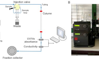
5. DNA Quality Check Using Gel Electrophoresis:
Following the extraction of DNA, it is crucial to assess its quality and integrity. This is usually accomplished through agarose gel electrophoresis, where DNA samples are placed into an agarose gel and exposed to an electric current. The negatively charged DNA fragments move towards the positive electrode, with smaller fragments traveling more quickly than larger ones.
Afterward, the gel is treated with ethidium bromide or SYBR Green, which binds to the DNA and emits fluorescence when exposed to UV light. Intact genomic DNA manifests as a clear band, while fragmented DNA presents as a smear. This procedure verifies the success of the DNA isolation process.
6. DNA Quantitation Using Fluorometric Method (QUBIT):
To achieve more precise and sensitive quantification of DNA, the Qubit fluorometer is utilized. This technique is highly selective, depending on DNA-binding dyes such as those found in the Qubit dsDNA Assay Kit. It enables accurate measurement of DNA, particularly at low concentrations, which is essential for subsequent processes like PCR.
Fluorometric methods, in contrast to spectrophotometry, are less susceptible to interference from contaminants like proteins or phenol, resulting in more reliable outcomes.
7. PCR Amplification Using Gene-Specific Primers:
The Polymerase Chain Reaction (PCR) is employed to amplify a particular gene of interest from bacterial genomic DNA. Primers specific to the gene are created based on the known sequences of the target gene.
Steps:
1. A PCR mixture is assembled, consisting of DNA template, primers, dNTPs, buffer, and DNA polymerase.
2. The mixture undergoes thermal cycling, which encompasses denaturation (generally at 94-98°C), annealing (ranging from 50-65°C), and extension (at 72°C).
3. The amplification is monitored by observing the increase in the size of the product.
A successful PCR yields a distinct band that corresponds to the gene of interest, which will be further analyzed in subsequent steps.
8. PCR Product Gel Extraction and Purification:
Following PCR amplification, the resulting product is subjected to agarose gel electrophoresis to verify the amplification's size and specificity. A gel extraction kit is utilized to cut out the targeted band from the gel, allowing for the purification of DNA from the agarose slice.
The procedure for gel extraction generally involves melting the gel slice, attaching the DNA to a column, and then eluting the purified DNA using a buffer. The purified PCR product is subsequently prepared for sequencing.
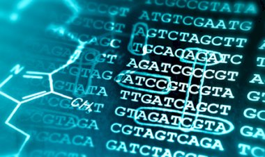
9. Gene Identification Using Sanger Sequencing:
The final phase in characterizing a gene involves determining its nucleotide sequence. Sanger sequencing, often referred to as the chain termination technique, is commonly employed for sequencing small segments of DNA. In this technique:
1. The purified PCR product is combined with a sequencing primer and a reaction mixture that includes dNTPs and fluorescently labeled dideoxynucleotides.
2. The DNA is then processed using a DNA sequencer. The resulting sequences are examined to accurately identify the nucleotide composition of the amplified gene.
3. Comparing this information with existing gene databases (such as GenBank) enables researchers to verify the gene's identity and its possible functions.
Training Programs at IBRI Noida
For those aspiring to build a career in Molecular Biology, the Indian Biological Sciences and Research Institute (IBRI) offers an excellent platform. IBRI provides a variety of courses and certifications that integrate both theoretical knowledge and hands-on training. Whether you're a beginner or an experienced professional looking to refine your expertise, IBRI's comprehensive programs ensure you gain practical experience in molecular biology techniques, experimental methodologies, and data analysis.
IBRI also provides specialized training in Bioinformatics, including In-silico Drug Designing, NGS, and Genomics & Proteomics analysis, equipping participants with industry-relevant skills.
These programs, led by experienced faculty, include practical training and certification, enhancing professional credentials.
Conclusion:
Molecular biology is transforming science, healthcare, and industry worldwide, driving advancements in genetic research and biotechnology. One such crucial aspect is the identification and characterization of genes from bacterial colonies, a complex process requiring a blend of laboratory techniques and analytical expertise. It requires precision in DNA extraction, amplification, sequencing, and data interpretation to ensure accurate results. Institutions like IBRI Noida are instrumental in offering the training and resources needed to develop the skills essential for these scientific investigations, helping researchers stay updated with the latest advancements in molecular biology.
Explore IBRI’s programs today to learn more about molecular biology trainings and unlock new career opportunities and stay ahead in the life sciences industry.
You can contact us at - Email: info@ibri.org.in or call at: (+91) 9999509892, 9971441910